Are you in need of CE credits? If so, check out our peer-reviewed, self-study CE courses here.
Test Your Saliva and Salivary Gland Knowledge
1. A healthy person produces about 600 mL of saliva per day, and this production increases during sleep.
It is estimated that a healthy person produces about 600 mL of saliva per day.1 Saliva production follows a circadian rhythm regulated by the suprachiasmatic nucleus in the hypothalamus, the body's central clock.2 Salivary flow rates are highest during the day and significantly decrease at night, approaching nearly zero during sleep.1,2
The circadian rhythm of the salivary glands plays a role in regulating food intake and immune system function, as it influences both saliva flow and its ionic composition. Disruptions to the circadian rhythm, such as sleep disturbance or insufficient sleep, can lead to inadequate saliva production and changes in its composition, increasing the risk of dental caries.2
References
1. Vila, T., Rizk, A.M., Sultan, A.S., Jabra-Rizk, M.A. The Power of Saliva: Antimicrobial and Beyond. PLoS Pathog. 2019; 15(11): e1008058. https://pmc.ncbi.nlm.nih.gov/articles/PMC6855406/
2. Kurtović, A., Talapko, J., Bekić, S., Škrlec, I. The Relationship between Sleep, Chronotype, and Dental Caries - A Narrative Review. Clocks Sleep. 2023; 5(2): 295-312. https://pmc.ncbi.nlm.nih.gov/articles/PMC10204555/
2. Which salivary glands begin to form first during prenatal development?
The parotid glands are the first to form, typically between 4 and 6 weeks during prenatal development. The submandibular glands follow at around 6 weeks, and the sublingual and minor glands begin forming at approximately 8 weeks. The parotid gland originates from the ectoderm, while the submandibular and sublingual glands are endodermal in origin.1
Gland development proceeds through distinct stages: bud and cord formation, branching of the cords, lobule formation, canalization, and cytodifferentiation. This process begins with a proliferation of oral epithelial cells that form buds, which then branch to create the final gland structure. The ducts and secretory end pieces complete their formation during the last two months of gestation. Glands continue to grow after birth, mainly due to an increase in acinar cell volume.1
Reference
1. Alhajj, M., Babos, M. (2023, July 24). Physiology, Salivation. StatPearls. https://www.ncbi.nlm.nih.gov/books/NBK542251/
3. Low salivary pH increases the risk of dental caries. A high salivary pH can contribute to increased erosion, calculus deposits, and black line stain.
The demineralization-remineralization process is an ongoing and dynamic process. Demineralization is the process of removing mineral ions from hydroxyapatite (HA) crystals via lowered pH, primarily caused by acidic attacks from dietary acids or oral bacteria. Conversely, remineralization is the process of restoring these mineral ions to the HA crystals. This process requires adequate calcium and phosphate ions, often supplied by saliva, and is significantly enhanced by the presence of fluoride. When the process is disrupted, there is a higher risk of developing dental caries.1
Erosion is a chemical process characterized by acids causing the dissolution of hard dental tissue, not including acids of bacterial origin. It is often a progressive loss of tooth mineral substances caused by either intrinsic or extrinsic factors. Examples of intrinsic factors include acid reflux and excessive vomiting, while extrinsic factors include medications and diet, such as the consumption of acidic beverages.1
An alkaline oral environment, while protective against some acids, promotes the formation of dental calculus. Certain oral ureolytic bacteria hydrolyze salivary urea to produce ammonia, which raises the salivary pH. This rise in pH increases the saturation of calcium phosphate in plaque, leading to the mineralization of plaque into calculus. Dental calculus then acts as a retentive surface and a reservoir for bacteria, which contribute to the initiation and progression of periodontal disease.2
Although the precise mechanism of black line stain remains unknown, studies suggest that high salivary pH may influence its formation.3
References
1. Abou Neel, E.A., Aljabo, A., Strange, A., et al. Demineralization-Remineralization Dynamics in Teeth and Bone. Int J Nanomedicine. 2016; 11: 4743-4763. https://www.ncbi.nlm.nih.gov/pmc/articles/PMC5034904/
2. D'souza, L.L., Lawande, S.A., Samuel, J., Wiseman Pinto, M.J. Effect of Salivary Urea, pH and Ureolytic Microflora on Dental Calculus Formation and its Correlation with Periodontal Status. J Oral Biol Craniofac Res. 2023; 13(1): 8-12. https://pmc.ncbi.nlm.nih.gov/articles/PMC9636048/
3. Al-Shareef, A., González-Martínez, R., Cortell-Ballester, I., et al. Current Perspective on Dental Black Stain of Bacterial Origin: A Narrative Review. Eur J Oral Sci. 2025; 133(3): e70007. https://onlinelibrary.wiley.com/doi/10.1111/eos.70007
4. Which salivary glands produce the greatest total volume of saliva in an unstimulated state?
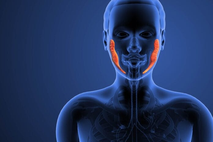
The submandibular glands produce the greatest total volume of saliva (approximately 65%–70%) in an unstimulated state.1,2 The remaining volume is contributed by the other major and minor salivary glands, with the parotid glands producing about 20%, the sublingual glands about 5%, and the minor salivary glands about 10%.2 While the submandibular glands produce the most saliva in an unstimulated state, the parotid glands produce more than 50% of saliva when stimulated.1
References
1. Grewal, J.S., Jamal, Z., Ryan, J. (2022, December 11). Anatomy, Head and Neck, Submandibular Gland. StatPearls. https://www.ncbi.nlm.nih.gov/books/NBK542272/
2. Vila, T., Rizk, A.M., Sultan, A.S., Jabra-Rizk, M.A. The Power of Saliva: Antimicrobial and Beyond. PLoS Pathog. 2019; 15(11): e1008058. https://pmc.ncbi.nlm.nih.gov/articles/PMC6855406/
5. A previously unidentified set of salivary glands was discovered in 2020.
In 2020, a previously unidentified set of salivary glands was discovered. The discovery was made during research on patients who had prostate or paraurethral gland cancer using a molecular imaging modality called positron emission tomography/computed tomography with radio-labelled ligands to the prostate-specific membrane antigen (PSMA PET/CT). Salivary glands are sensitive to the PSMA ligand, which makes them visible on these scans. These newly identified glands were named "tubarial glands" because of their location near the torus tubarius in the posterolateral wall of the nasopharynx.1
Their discovery is significant because it is believed they are associated with side effects like xerostomia and dysphagia in patients undergoing radiation therapy for head and neck cancers.1
Reference
1. Valstar, M.H., de Bakker, B.S., Steenbakkers, R.J.H.M., et al. The Tubarial Salivary Glands: A Potential New Organ at Risk for Radiotherapy. Radiother Oncol. 2021; 154: 292-298. https://www.thegreenjournal.com/article/S0167-8140(20)30809-4/fulltext


