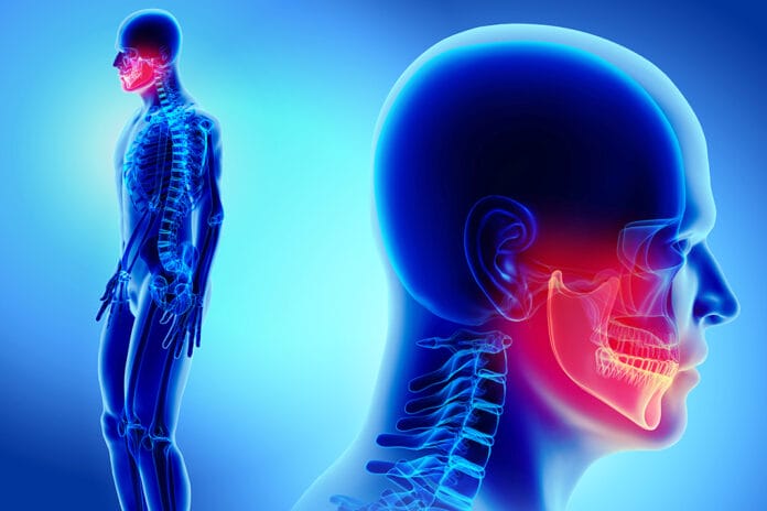
With health conditions becoming more complex, the dental hygienist’s review of health histories has become more confusing. Many conditions sound and seem the same but have slight variances that make the difference, such as within the “osteo” group of conditions. While “osteo” means relating to bones, these conditions affect not only the skeleton but may also affect oral health.
The human skeleton completely regenerates every 10 years. Bone is constantly resorbing through osteoclasts and forming through osteoblasts. Many “osteo” defects are caused by the osteoclasts’ insufficient production or defective function. The maintenance of bone depends on bone remodeling and the balance between the osteoclasts and osteoblasts. When there is an imbalance, a variety of bone conditions can occur.1
Osteopenia
Osteopenia (osteo = bone) (penia = lacking) is a decrease in bone mineral density that weakens bones. It’s the imbalance between bone resorption and formation favoring resorption, resulting in demineralization of the bone. It ranks between healthy bone and osteoporosis and can lead to osteoporosis if not maintained.2
The condition is diagnosed by the measure of T-score, which refers to the density of an individual’s bones compared with the average bone density of a young person of the same sex. Normal T-score is -1 and above. Osteopenia has a T-score between -1 and -2.5, and osteoporosis is -2.5 and lower. The lower scores for osteopenia are more common in women over age 50.2
Though current research at this time shows no correlation between the lowered density of skeletal bone seen in osteopenia and a reduction in periodontal clinical attachment loss, osteopenia is worth mentioning as it increases the risk of osteoporosis.3
- Symptoms: Usually none; some may experience bone pain or weakness, trouble walking, bending or twisting, loss of height, curved or stooped spine, brittle nails, or bone fracture from a minor event2
- Bones most commonly affected: Fractures occur most often in the hip, wrist, forearm, humerus, and spine4
- Causations or risks: Unhealthy lifestyle, medications, poor nutrition, hormonal changes, smoking, heavy alcohol use, diabetes, sedentary lifestyle, hereditary, age, female, untreated celiac disease, overactive thyroid, and chemotherapy2
- Common treatments: Calcium, exercise, healthy diet, vitamin D2
Paget’s Disease
Paget’s disease is a chronic condition that affects the bone remodeling process when the bone breaks down, and the rebuilding process increases. This can cause unusual and disorganized bone structure and promotes softer, weaker, or larger bones, making them susceptible to complications such as bending or fractures.5
When Paget’s disease affects the jaw, the risks to oral health can be substantial. The loss of alveolar bone causes mobility or loss of teeth, overgrowth of bone causes spreading of teeth and malocclusion, and improper fit of dentures. More complications include root resorption, increased cementum, excessive bleeding with extractions, and osteomyelitis.6
- Signs and Symptoms: Hearing loss if disease occurs in the temporal bone; cardiovascular, neurologic, and metabolic complications may occur; osteoarthritis in adjacent joints is common, with aching pain caused by small fractures or nerve compressions; visible deformities of enlarged skulls or curvature of the femur
- Bones commonly affected: Spine, skull, pelvis, and legs
- Causations or risks: Hereditary, age, European descent
- Common treatments: Bisphosphonates, surgery, exercise, and nutritional diet6
Osteopetrosis
Osteopetrosis (osteo = bone) (petrosis = hard), which is often referred to as stone bone, is a rare inherited skeletal disorder of increased bone density and abnormal bone growth during the formation of bone. Instead of the normal process of old bone breaking down and new bone forming, only new bone is formed, causing thick, dense bones to develop improperly. Bones can become overly dense and cause misshapen bones that are large, brittle, and easy to fracture.1
The increased size of bone invades the bone marrow area, resulting in less space for bone marrow. This leads to a decrease in immune cells and cells that carry oxygen and control bleeding.1
Osteopetrosis can be noticeable from birth to adulthood, with complications ranging from mild and only discovered through incidental findings on a radiograph to severe. Depending on the severity and type of osteopetrosis, it can cause nasal congestion from narrow sinus cavities, vision and hearing complications, and a decrease in blood cells, which may lead to infection and anemia, chronic bone pain, and osteomyelitis.1
Malignant infantile osteopetrosis (apparent from birth) frequently shortens life expectancy if left untreated within the first 10 years. This form of osteopetrosis has the most severe symptoms and defects, including hydrocephalus, liver and spleen enlargement, and pancytopenia, a deficiency of all blood cells.1
In adult-onset osteopetrosis (late childhood to adulthood), symptoms are milder with gradual deterioration.1
In terms of oral health, osteopetrosis causes changes in the skull and jaw, leading to delayed tooth eruption, severe dental caries, micrognathia, mandibular prognathism, osteomyelitis of the jaw, dental anomalies, deciduous retention, malformation of teeth crowns, abscesses, and facial paralysis.1
- Signs and Symptoms: Fractures, decreased blood cell production, frequent dental infections, inhibited growth, eating difficulties, and loss of cranial nerve function, which may cause blindness, deafness, facial nerve paralysis
- Bones commonly affected: Skull, ribs, long bones, and vertebrae
- Causations or risks: Inherited adult type: autosomal dominant; infantile type: autosomal recessive, and x-linked recessive
- Common treatments: In specific cases of infantile type osteopetrosis, hematopoietic stem cell transplantation (HSCT) and gamma-1b injections may be appropriate depending upon genetic studies; if HSCT treatment is deemed inappropriate, corticosteroids may be considered, but there isn’t good evidence supporting their routine use; good nutrition with calcium and vitamin D is important as treatment tends to be supportive based on symptoms1
Osteomyelitis
Osteomyelitis (osteo = bone) (myelitis = inflammation) occurs when bacteria or fungi from infected surrounding tissue enter the bone via the bloodstream. Staphylococcus aureus is a common bacterium with osteomyelitis.7 This condition can lead to osteonecrosis if the infection reduces blood flow.6
Osteomyelitis may occur at any age, including young children. It appears more often in men than women.8
The oral health discussion centers on osteomyelitis in the mandible from a microbial infection that reaches the bone, arising from periodontal lesions, abscessed teeth, root canal therapy, or traumatic injuries. Recent surgeries prior to dental work can increase the risk of developing an infection in the jaw. Those with decreased immune system function are more vulnerable to infection during the healing process.9
On a panoramic radiograph, osteomyelitis will present with an ill-defined trabecular bone pattern with areas of radiolucencies known as “moth-eaten.” It takes six to 12 months for the bone to remodel gradually for successful treatment.9
- Symptoms: Drainage of pus, fever, pain, swelling, tender to touch, limited and painful movement, fatigue, head and neck pain, deep-seated throbbing pain in the jaw or forehead, halitosis, difficulty eating, and numbness or paresthesia of lower lip
- Bones affected: The adjacent ends of long bones, spine, and mandible more so than maxillary, as it is more vascular
- Causations or risks: Artificial joints, blood infections, diabetes, especially a diabetes-related foot ulcer, metal implants in bones, pressure injuries, compromised immune system, alcohol addictions, recent broken bones, and traumatic injuries or wounds
- Common treatments: Antibiotics, antifungals, pain relievers, and bone surgery9,10
Osteoporosis
Osteoporosis (osteo = bone) (porosis = holes) is a skeletal disorder featuring compromised bone strength and decreased bone mass and mineral density. The process of bone breaking down becomes faster than the bone being rebuilt. This imbalance causes thinner, weaker bones as well as a loss of bone integrity, increasing the risk of fracturing. It affects more women, especially through menopause, as estrogen is lost and more bone is removed than laid down.11
There’s a modest correlation between bone mineral density between the maxillary, mandible, and alveolar bone with overall skeletal density. Dense mandibular alveolar trabecular patterns are strongly correlated with higher skeletal bone mineral density.6 If osteoporosis is present, it may show in dental radiographs, including panoramic images. The morphology of the inferior mandibular cortex and architecture of trabecular bone may present altered or deteriorated.12
Further oral implications for patients with osteoporosis may include the loss of periodontal attachment, resorption of both condyles and perhaps the TMJ, and a decrease in the height of alveolar bone due to resorption.13
Patients with osteoporosis have elevated systemic levels of proinflammatory cytokines, which are osteoclastogenic bone resorption-inducing cytokines that cause low bone density. Circulatory inflammatory cytokines from periodontitis can impact bone remodeling.14
- Signs and Symptoms: Bone fractures, stooped posture, loss of height, back pain
- Bones commonly affected: Spine, hips, wrists, forearms, and jaw
- Causations or risks: Age, hormones, family history, smoking, poor nutrition, low body weight or smaller frame, alcohol use, chemotherapy drugs, sedentary lifestyle
- Common treatments: Bisphosphonates, weighted exercises, calcium, vitamin D15
Osteonecrosis
Osteonecrosis (osteo = bone) (necrosis = death) is considered avascular or aseptic necrosis. It occurs when the blood flow is disrupted, leading to bone tissue death. The bone breaks down, and the joint collapses. This most commonly happens in the shoulders, knees, and hips, and, within dentistry, the mandible. The jaw regenerates more than other bones, leaving it more receptive to anti-resorptive medications as they target and inhibit osteoclasts.16
Medication-related osteonecrosis of the jaw (MRONJ) is mostly derived from the bisphosphonate class of drugs. These medications may be taken in high doses for bone cancer treatment or cancer that has spread to the bones. These medications can also be taken in lower doses for severely decreased bone density, such as osteoporosis or osteogenesis imperfecta. These medications can prevent the jaw from healing properly and expose the bone.17
Other contributing factors of patients taking these medications are active periodontitis, smoking, diabetes, ill-fitting dentures, and older age. Symptoms of MRONJ are gingiva that does not heal, lack of tissue around the bone, numbness or heaviness in the jaw, tooth mobility, and infection of the gingiva or jaw with pain or swelling.17
Osteonecrosis of the jaw (ONJ) is considered avascular necrosis, meaning blood supply is lost to the region. The condition may occur after dental extractions, bone grafting, or insertion of dental implants. It occurs when the bone doesn’t heal and is left uncovered. Exposed bone does not receive blood flow, and the cells die.18
The condition is not to be mistaken for osteoradionecrosis, which occurs after radiation therapy for head and neck cancers. Radiation therapy to the head and neck can destroy blood vessels carrying blood to the bones. The increased risk of osteoradionecrosis emerges from invasive dental procedures after radiation therapy.18
When osteonecrosis stems from an injury, it’s considered traumatic, while all other necrosis cases are considered nontraumatic or atraumatic.19
- Symptoms: Pain is often related to exposed bone, soft tissue swelling and draining, infection, mobility or loss of teeth, jawbone fractures, and periodontal infections
- Bones most commonly affected: Jaw, the upper part of the humerus at the ball and joint of the shoulder, the upper part of the femur at the ball and hip socket, and the lower femur at the knee
- Causations or risks: Cancer treatments, history of medication use including bisphosphonates, dental trauma, injury, blood disorders, rheumatoid arthritis, decompression sickness, gout, surgery, or infections
- Common treatments: Rinses, antibiotics, oral analgesics, and surgical debridement with monitoring thereafter16,19
Due to the increased risk of osteonecrosis in patients taking bisphosphonates, thoroughly reviewing medical history and discussing these medications with patients is imperative. Most benefits seen in patients taking bisphosphonates can be observed in the first five years. Therefore, it is generally recommended that patients have regular follow-up visits with the prescribing physician to determine if or when the benefit of the medication no longer outweighs the risk of developing complications, such as osteonecrosis.20
With the prolonged embedding of bisphosphonates in the bone tissue, the risks of these medications can last for years. Therefore, knowing the history of osteoporosis medication use is important.21
Osteoporosis Medications
- Alendronate (Fosamax, Binostso): Prevents and treats postmenopausal osteoporosis and treats osteoporosis in men. It also treats secondary osteoporosis in those taking glucocorticoids. Taken orally daily or weekly.
- Ibandronate (Boniva): Treats postmenopausal women only. Taken orally monthly or intravenously once every three months.
- Risedronate (Actonel, Atelvia): Prevents and treats osteoporosis caused by menopause, hypogonadism, and prolonged steroid use. Taken orally in varying doses depending on sex, age, and underlying cause of osteoporosis, usually daily, weekly, or monthly.
- Zoledronic acid (Reclast, Aclastia, Zometa): Prevents and treats osteoporosis from menopause, hypogonadism, and steroid use. Taken intravenously once a year.
- Denosumab (Prolia): A monoclonal antibody that works by preventing the development of osteoclasts. It is used in patients who have osteoporosis and are at high risk for fractures. Taken by injection every six months. Similar to bisphosphonates, it increases the risk of osteonecrosis of the jaw, infections, jaw pain, and tooth mobility.20
Osteochondroma
Osteochondroma (osteo = bone) (chondro = cartilage) (oma = tumor) is the overgrowth of cartilage and bone, causing a benign tumor at the end of the bone near the growth plate. It mainly occurs in childhood or adolescence when the bones are growing.22
In normal bone growth, the bone occurs from the growth plate and hardens into solid bone when the person is fully grown. In osteochondroma, there’s an outgrowth of the growth plate consisting of cartilage and bone. During the growth period, osteochondroma becomes more prominent; however, tumors don’t develop or enlarge after puberty.22
Osteochondromas may form as solitary (affecting males and females equally) or multiple (affecting males more). Solitary osteochondroma is considered the most common benign bone tumor, accounting for about 20-50% of benign bone tumors and 9% of all bone tumors. Instead of bone growing in line with the growth plates, it grows out from it, mainly at the knee, hip, and shoulder joints.22
Hereditary multiple osteochondromas (HMO) are developed due to a rare genetic disorder affecting one of the EXT genes. Signs and symptoms may include pain, decreased range of motion, nerve impingement, deformities, differences in limb length, short stature, and fractures. The location of the HMO will vary from person to person. Most HMOs are benign; however, around 2-5% of people will experience HMOs becoming malignant in adulthood. About 96% of females with a mutation in one of the EXT genes will develop osteochondromas, while 100% of males with the mutation will develop HMO.23
When osteochondroma occurs in the jaw, it can cause enlargement or abnormal growth, leading to facial deformities, masticatory and occlusive symptoms, and pain. Only about 1% occur in the head and neck region and slightly affects women more than men. Within the jaw, older individuals are affected more often; younger individuals are affected more in the long bones.24
- Symptoms: Commonly none, but if there are some, they include one leg/arm longer than the other, soreness of nearby muscle, pressure or irritation with activity, shorter than normal height for age, and a hard, painless, immovable mass
- Bones commonly affected: Long bones of the legs, pelvis, and shoulder blade, less common in craniofacial regions but can happen in the mandible and condyle and coronoid tip
- Causations or risks: Definitive causes are unknown but thought to be caused by the EXT1 gene; bone cancers may occur if bone cells grow uncontrollably
- Common treatments: Nonsurgical with observation or surgical by removing the tumors22
Osteosarcoma
Osteosarcoma is a primary malignant cancer of long bones. It is the second most common malignant condition of the bone, accounting for about 15-20% of all primary bone tumors. It occurs commonly in the distal femur, proximal tibia, and humerus. The condition is more common in males than females, especially in young adults, peaking in frequency during adolescent growth spurts. Then another small peak of osteosarcoma happens in adults over 50.23
Osteosarcoma represents up to 10% of the cases within the jaw and accounts for 23% of the primary malignant tumors of the head and neck. Within the jaw, it appears more frequently in the mandible (specifically the ramus) than the maxilla, specifically the canine and premolar regions.26
Within dentistry, primary symptoms include pain, swelling, paresthesia, and ulceration. Osteosarcomas have a better prognosis within the jaw than long bone osteosarcoma cases, with an estimated cure prognosis of about 70%. The prognosis is better in the mandible than the maxilla, as resection and obtaining negative surgical margins are more successful because of less anatomical complexity. Paget’s disease can be a predisposed factor to the development of osteosarcomas.26
Ewing Sarcoma
Ewing sarcoma develops when the DNA in certain cells changes. These cells become abnormal and attack healthy tissue. Ewing sarcoma is an aggressive form of cancer and quickly metastasizes, commonly to the bones and lungs.25 Ewing sarcoma accounts for 6% of all malignant bone tumors, with approximately 4% occurring in the head and neck region and 1% in the jaw.28
When Ewing sarcoma occurs in the mandible, it’s usually located in the posterior regions, including the ramus. Since Ewing carcinoma tends to metastasize to other bones, it will likely metastasize to the jaw from other skeletal regions.28
Osteoarthritis
Osteoarthritis (osteo = bone) (arthritis = joint inflammation) is also referred to as degenerative joint disease or “wear and tear” arthritis. It affects the body’s joints through the gradual degradation of cartilage that covers the surface of the joints. Osteoarthritis can cause a change in the shape of the bones.6
Osteoarthritis can decrease physical, mental, and general health, as activity, energy, and sleep can be limited while pain increases.6
Osteoarthritis in the temporomandibular joints occurs when the cartilage disk at the joint between the skull and mandible degenerates, and this can cause pain and an altered occlusion. With severe osteoarthritis, the patient has limited opening of the mouth.6
Oral home care may be affected in patients with osteoarthritis due to dexterity issues and the inability to remove plaque and food thoroughly. With limited mobility, patients may skip routine dental visits, thus causing more emergencies, extractions, periodontal disease, and restorative and prosthodontic treatment.6
- Symptoms: Pain and stiffness accompanied by loss of function, decreased flexibility, and swelling
- Joints commonly affected: Knees, hands, feet, hips, spine, and temporomandibular joints
- Causations or risks: Age, wear and tear, stress from weight, increased joint instability, laxity, vulnerability, mechanical insults, and decreased resilience and reparative capacity of cartilage
- Common treatments: Increased physical activity, weight loss, physical therapy, corticosteroids, and NSAIDS; corticosteroids may suppress immune function, leading to delayed wound healing, fungal infections, and prolonged bleeding6,29
Osteomalacia
Osteomalacia (osteo = bone) (malacia = tissue softening) occurs when the bones do not normally mineralize after forming. Prolonged vitamin D deficiency and its effects on calcium serum levels cause impaired bone metabolism, leading to incomplete mineralization of bones.30
Physical signs of osteomalacia include deformities such as lordosis with a waddling gait. Bones do not regain their original shape after deformation.30
Risk factors for osteomalacia include European descent, dark skin, limited sun exposure, low socioeconomic status, and poor diet.30
Osteomalacia should also not be confused with osteoporosis, which is the weakening of living bone already formed and remodeled. Osteomalacia is a problem with the forming of bones.15,30
Osteomalacia should not be confused with rickets in children, which affects the growth plates of the bones in the developmental process. However, osteomalacia and rickets often occur concurrently.5,30 When rickets and osteomalacia are concurrent, children may have premature tooth loss and need orthodontic treatment.31
- Symptoms: Bone fractures without injuries, muscle weakness, bone pain starting in the lower back and thighs and spreading to the arms and ribs, spasms or cramps of the hands and feet, trouble standing up, using stairs, and walking
- Bones most commonly affected: Skeletal of long bones and spine, hips, lower back, and feet
- Causations or risks: Vitamin D deficiency, low blood calcium, kidney conditions, liver disease medications, untreated celiac disease, stomach surgery
- Common treatments: Correcting underlying nutrient deficiency, weight-bearing exercises, and maintaining a healthy weight and diet31
Osteogenesis Imperfecta
Osteogenesis imperfecta (osteo = bone) (genesis = formed) (imperfecta = faulty), which is also called brittle bone disease, is a genetic bone disorder from birth. People with this disease are born with fragile bones that easily fracture for no reason. Patients with osteogenesis imperfecta have a change in their genes that interferes with the production of collagen, leading to weak bones.32
Problems from osteogenesis imperfecta range from mild to severe, with eight different types of the disease. It affects men and women the same.33
Osteogenesis imperfecta may affect the growth of the jaws and the teeth. Some patients with osteogenesis imperfecta also have dentinogenesis imperfecta, a disorder of tooth development that presents as brittle, opalescent, misshapen teeth, which may break or chip easily. Dentinogenesis imperfecta can be diagnosed with the first baby tooth appearing as gray, bluish, or brown. Radiographically, crowns will appear bulbous, and roots will be shorter and more slender than normal teeth.33,34
In the type III variance of osteogenesis imperfecta, the narrowing of the cervical portion of the tooth over-accentuates a bulbous appearance of the crown, although the crowns are normal size. The roots are short, with partial or complete obliteration of the pulp chambers and canals. Primary teeth have larger pulp chambers until the dentin is formed.34
Regular dental appointments are important as orthodontics, veneers, crowning or bridges of the teeth, surgeries, partials, or dentures may be needed.
- Signs and Symptoms: Broken bones, bone deformities with bowing of the legs, barrel-shaped chest, curved spine, triangle-shaped face, loose joints, muscle weakness, bruises easily, blue or gray discoloration of the white of the eye, hearing loss in early adulthood, respiratory infections, and heart problems, such as poor heart valve function
- Bones commonly affected: Long bones, spine, lower back, hips, pelvis, legs, arms, and ribs
- Causations or risks: Genetics with up to 90% caused by a dominant mutation in a gene coding for type 1 collagen
- Common treatments; No cure; orthopedic treatment, physical therapy, weight management, and avoidance of fractures and infections; bisphosphonate medicines reduce the remodeling rate of bones but could lead to osteonecrosis 33,34
Conclusion
Dental recalls are important for patients not only to maintain oral health but also for bone health. As dental professionals, if we notice any abnormal bone presentation after ruling out oral disease, discuss these findings with your patient and refer them to their primary care physician for further testing and follow-up care. Or vice versa, if a patient starts a new medication or lists a new condition related to bone health, we know to take caution during certain dental treatments and their related risks.
Before you leave, check out the Today’s RDH self-study CE courses. All courses are peer-reviewed and non-sponsored to focus solely on pure education. Click here now.
Listen to the Today’s RDH Dental Hygiene Podcast Below:
References
- Osteopetrosis. (2023, November 20). National Organization for Rare Disorders. https://rarediseases.org/rare-diseases/osteopetrosis/
- McDowell, S. (2023, March 15). What Is Osteopenia? https://www.healthline.com/health/osteopenia
- Zamani, S., Kiany, F., Khojastepour, L., et al. Evaluation of the Association Between Osteoporosis and Periodontitis in Postmenopausal Women: A Clinical and Radiographic Study. Dent Res J. 2022; 19: 41. https://pdfs.semanticscholar.org/3512/a7203e9b0ac314fcbbf627b71083d6546235.pdf
- Varacallo, M., Seaman, T.J., Jandu, J.S., et al. (2023, August 4). Osteopenia. StatPearls. https://www.ncbi.nlm.nih.gov/books/NBK499878/
- Santhakumar, S. (2023, February 16). What to Know About Bone Disease. Medical News Today. https://www.medicalnewstoday.com/articles/bone-diseases#conditions
- Kelsey, J.L., Lamster, I.B. Influence of Musculoskeletal Conditions on Oral Health Among Older Adults. Am J Public Health. 2008; 98(7): 1177-1183. https://ajph.aphapublications.org/doi/full/10.2105/AJPH.2007.129429
- Osteomyelitis. (n.d.). Cleveland Clinic. https://my.clevelandclinic.org/health/diseases/9495-osteomyelitis
- Kremers, H.M., Nwojo, M E., Ransom, J.E., et al. Trends in the Epidemiology of Osteomyelitis: A Population-based Study, 1969 to 2009.The Journal of Bone and Joint Surgery. 2015; 97(10): 837-845. https://doi.org/10.2106/JBJS.N.01350
- Auluck, A. How do I Manage a Patient with Osteomyelitis? Journal of Canadian Dental Association. 2016; 82: 13. https://jcda.ca/g13
- Kusuyama, Y., Matsumoto, K., Okada, S., et al. Rapidly Progressing Osteomyelitis of the Mandible. Case Reports in Dentistry. 2013; 2013: 249615. https://www.ncbi.nlm.nih.gov/pmc/articles/PMC3855945/
- U.S. Department of Health and Human Services. (2004). Basics of Bone in Health and Disease (pp. 16-36). In Bone Health and Osteoporosis: A Report of the Surgeon General. CreateSpace Independent Publishing Platform. https://www.ncbi.nlm.nih.gov/books/NBK45504/
- Graham, J. Detecting Low Bone Mineral Density from Dental Radiographs: A Mini-review. Clinical Cases in Mineral and Bone Metabolism. 2015; 12(2): 178-182. https://www.ncbi.nlm.nih.gov/pmc/articles/PMC4625777/
- Bhatnagar, S., Krishnamurthy, V., Pagare, S.S. Diagnostic Efficacy of Panoramic Radiography in Detection of Osteoporosis in Post-Menopausal Women with Low Bone Mineral Density. J Clin Imaging Sci. 2013; 3: 23. https://www.ncbi.nlm.nih.gov/pmc/articles/PMC3690705/
- Wang, C.J., McCauley, L.K. Osteoporosis and Periodontitis. Curr Osteoporos Rep. 2016; 14(6): 284-291. https://www.ncbi.nlm.nih.gov/pmc/articles/PMC5654540/
- Osteoporosis. (2022, December). National Institute of Arthritis and Musculoskeletal and Skin Diseases. https://www.niams.nih.gov/health-topics/osteoporosis
- Osteonecrosis of the Jaw. (2023, February). American College of Rheumatology. https://www.rheumatology.org/I-Am-A/Patient-Caregiver/Diseases-Conditions/Osteonecrosis-of-the-Jaw-ONJ
- Mark, A.M. What is MRONJ? Journal of the American Dental Association. 2021; 152(8): 710. https://jada.ada.org/article/S0002-8177(21)00310-X/fulltext
- Osteonecrosis of the Jaw (ONJ). (2022, September 7). Cleveland Clinic. https://my.clevelandclinic.org/health/diseases/24156-osteonecrosis-of-the-jaw
- Osteonecrosis. (n.d.). National Institute of Arthritis and Musculoskeletal and Skin Diseases. https://www.niams.nih.gov/health-topics/osteonecrosis
- Curtis, S. (2019, May 23). Osteoporosis Medications. Spine-Health. https://www.spine-health.com/conditions/osteoporosis/osteoporosis-medications
- Ott, S.M. Long-term Safety of Bisphosphonates.The Journal of Clinical Endocrinology and Metabolism. 2005; 90(3): 1897-1899. https://doi.org/10.1210/jc.2005-0057
- Tepelenis, K., Papathanakos, G., Kitsouli, A., et al. Osteochondromas: An Updated Review of Epidemiology, Pathogenesis, Clinical Presentation, Radiological Features and Treatment Options. In Vivo. 2021; 35(2): 681-691. https://doi.org/10.21873/invivo.12308
- Hereditary Multiple Osteochondromas. (n.d.). National Center for Advancing Translational Sciences. https://rarediseases.info.nih.gov/diseases/7035/hereditary-multiple-osteochondromas
- Kishore, D.N., Kumar, H.R.S., Umashankara, K.V., et al. Osteochondroma of the Mandible: A Rare Case Report. Case Reports in Pathology. 2013; 2013. https://doi.org/10.1155/2013/167862
- Osteosarcoma. (n.d.). Physiopedia. https://www.physio-pedia.com/Osteosarcoma
- Nthumba, P.M. Osteosarcoma of the Jaws: A Review of Literature and a Case Report on Synchronous Multicentric Osteosarcomas. World J Surg Onc. 2012; 10(240). https://doi.org/10.1186/1477-7819-10-240
- Ewing Sarcoma. (n.d.) Cleveland Clinic. https://my.clevelandclinic.org/health/diseases/21752-ewings-sarcoma
- Krishna, K.B., Thomas. V., Kattoor, J., Kusumakumari, P. A Radiological Review of Ewing’s Sarcoma of Mandible: A Case Report with One Year Follow-up. Int J Clin Pediatr Dent. 2013; 6(2): 109-114. https://www.ncbi.nlm.nih.gov/pmc/articles/PMC4086594/
- Zhao-Fleming, H., Hand, A., Zhang, K., et al. Effect of Non-steroidal Anti-inflammatory Drugs on Post-surgical Complications against the Backdrop of the Opioid Crisis. Burns and Trauma. 2018; 6: 25. https://doi.org/10.1186/s41038-018-0128-x
- Zimmerman L, McKeon B. (2024, September 2). Osteomalacia. StatPearls. https://www.ncbi.nlm.nih.gov/books/NBK551616/
- Case-Lo, C. (2024, April 17). Osteomalacia. Healthline. https://www.healthline.com/health/osteomalacia
- Osteogenesis Imperfecta Overview. (n.d.). National Institute of Arthritis and Musculoskeletal and Skin Diseases. https://www.bones.nih.gov/health-info/bone/osteogenesis-imperfecta/overview
- 33. Dentinogenesis Impefecta. (n.d.). National Library of Medicine. https://medlineplus.gov/genetics/condition/dentinogenesis-imperfecta/
- Vammi, S., Bukyya, J.L., Ck, A.A., et al. Genetic Disorders of Bone or Osteodystrophies of Jaws – A Review. Glob Med Genet. 2021; 8(2): 41-50.


