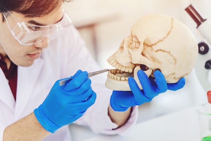
The mouth is one of the most unique features of the body ‒ most valuable in the basic survival of speaking and eating. The mouth is also very valuable in providing identification. Fingerprints have distinct attributes, and so do the lip, tongue, teeth, and rugae. These prints can identify people in uncertain situations such as crimes, mass disasters, missing persons, and biometrics.
Lips
Lips have many purposes, from emotional expressions with smiling to sensory, sexuality and sensuality, and to the full function of talking, eating, drinking, and swallowing.1 They also can be a source of identification. The fissures such as wrinkles and grooves on the lips have their own special marks to form patterns referred to as lip prints.
Patterns on the lips are seen as early as the sixth week in the womb and stay uniform throughout life. Lip prints stay in contact during minor alterations of trauma, inflammation, and sores. Although major trauma, scarring, and surgical treatment for pathosis may alter the patterns and morphology with the size and shape of the lips, the prints will resume their former pattern post-recovery (except with burns).2
The study of lip prints is called cheiloscopy. Dr. Martin Santos recognized that lip patterns could be used for personal identification in 1960, and he developed a system to determine types of lip grooves. The four different classifications are straight, curved, angled, and sine-shaped lines.
In 1967, Kazuo Suzuki, a Japanese scientist, developed a classification of the lips’ measurement, use, and color. Later in 1970, Suzuki and Yasuo Tsuchihashi produced a method of classification of six lip prints:3,4
- Type I: A clear-cut groove running vertically throughout the lip from the border of the lip to just inside the lip (dominant in females)
- Type I’: Partial-length groove of type 1 (dominant in females)
- Type II: Branched grooves (dominant in females)
- Type III: Intersected grooves (dominant in males)
- Type IV: Reticular patterns (dominant in males)
- Type V: Other patterns and grooves that are morphologically differentiated dominant in females and males3,4
The main lip structures used for identification are the size of the lips by measuring the maximum vertical dimension between the vermillion border of both the upper and lower lips. The other is the shape of the oral fissure and upper and lower vermillion borders. For identification use, the Glogau-Klein points are very beneficial. This area is covered with wrinkles and grooves to form a distinct lip pattern. Lip thickness and position are also determined in lip prints and are expressed as thin, thick or very thick, medium, or mixed.2
Lip prints may be visible or nonvisible. The vermillion border of the lips contains minor salivary glands, and, from licking the lips with the tongue, latent prints can be discovered and used. A closed mouth exhibit defined grooves, while an open position shows the grooves less defined in capturing lip prints
Lip prints can occur on anything, including paper, tape, glass, clothing, cigarettes, utensils, and straws. The prints can be collected by several methods: chemical, powders, x-rays, lights, or magnifying lenses. Red lipstick and blotting on paper, tape, or a nonporous surface is also a credible source of lip print collection.3
Tongue
No two tongues are the same. Tongue prints used in biometrics are done by digitally scanning the tongue to gather important information of the shape and texture combined. The geometric shape and the physiological surface of the tongue stay consistent, making the individual, unique characteristics very difficult to manipulate or duplicate.
Tongue prints acquired from the dorsal and lateral borders are used in conjunction with other characteristic features such as surface and shape. The length, width, and thickness along with tongue textures of smooth, cracked, fissured, or scrotal determine tongue prints.
The key characteristics of the top surface of the tongue are the presence of central fissures, the number of fissures, and the depth of fissures. Grooves on the tongue may be single or multiple, shallow, or deep. Multiple shallow vertical fissures are more common in men while single deep vertical fissures are more common in women.5
Certain shape characteristics to determine tongue prints are ovoid, rectangular, triangular, square, and round. Other shape aspects are u-shaped, appearing in both men and women. A V-shaped tongue appears more often in women. The tip of the tongue may have a fibrous band or a mild or partial cleft. The shape is either bigger at the base and thinner at the tip or broad or thin throughout.
Tongue prints are obtained in a variety of ways. One such way is analysis through alginate impressions followed by a cast preparation to obtain the unique features of the tongue. Other methods are digital software, photographs, and videos.6
Teeth
Tooth prints are unique not only between people but also between teeth within the same mouth. Tooth prints are the term used to describe patterns of enamel rod ends, while the study of these patterns is called meloglyphics.
The basic structure of enamel is the enamel rods. Enamel doesn’t remodel once it’s lost as there’s no use for the ameloblasts after the tooth has matured and erupted. Each tooth contains millions of enamel rods, with the amount varying between teeth. A greater amount of enamel rods are in the thicker areas of the tooth, such as the cusps, and lesser amounts are in the thinner parts of the tooth, such as the cervical area. The size and diameter of the enamel rods are larger towards the outer surface of the enamel.
The shape of the enamel prisms to form the enamel rods consist of three main patterns:7
- Pattern 1 ‒ Circular
- Pattern 2 ‒ Aligned in parallel rows
- Pattern 3 ‒ Aligned in staggered rows where the tails lie between the heads, appearing like a keyhole
The patterns of the enamel rods create specific subpatterns that form a combination of patterns in each tooth to form their unique print. The subpatterns are wavy-branched, wavy-unbranched, linear-branched, linear-unbranched, whorl-open, whorl-closed, loop, and stem-like.
Conventionally, enamel rods are not laid in a straight path but rather intertwining to enforce high tensile strength. Enamel rods start from the dentinoenamel junction and branch to the outer enamel surface.
Generally, enamel rods lay perpendicular to the dentin surface. In primary teeth, the enamel rods are horizontal to the cervical and middle third of the teeth, become more oblique in the occlusal and incisal thirds, and lay vertical in the incisal edge and cusp tips. In permanent teeth, it’s similar to the primary teeth except in the cervical third, where the enamel rods deviate apically.7
Enamel rods are collected and evaluated through different techniques such as acid etching or the peel method. Frequent collection of these prints can be beneficial for personal identification, especially with firefighters, soldiers, divers, pilots, and frequent travelers. Due to wear and tear and enamel loss, updated prints periodically could be beneficial if this way of identification is ever needed.
Rugae
Rugae (the wrinkled or folded anatomy) is located on the anterior third of the palate behind the center of the maxillary incisors. This anatomy is individually unique, with the size, shape, and number, even including each side of the palate being distinctive. During the 12 to 14th week, the rugae form prenatally and maintain their shape throughout life. They will grow as the mouth grows, but they stay in the same position throughout life. Rugae seldom change shape with age and will reappear after trauma or surgical procedures (although in rare occasions, disease or trauma may alter the shape).
The study of rugae is called palatoscopy or palatal rugoscopy. Rugae prints can be an additional oral usage for identifying purposes, especially in edentulous individuals. A person’s rugae can be compared with present or older dentures. Analysis from maxillary impressions or photos is specific to evaluate the number of rugae, length of primary (5mm to 10mm), secondary (3mm to 5mm), and fragmentary (less than 3mm). More specific details are fusion and course of direction from the mid-palatine raphe and the predominant shape of rugae.
The rugae shapes include:5
- Straight rugae ‒ Follows a straight or linear line from the origin to termination
- Curved rugae ‒ Is crescent or a slight bend at the origin or termination
- Wavy rugae ‒ Is curved or slightly curved at the origin or termination in the form of a wave
- Circular rugae ‒ Has a definite circular ring
- Unification (diverging) rugae ‒ Is two rugae with a common origin but separate terminations
- Unification (converging) rugae ‒ Is two rugae with separate origins and uniting laterally at the termination
The mouth is interesting and provides unique characteristics. These characteristics can be used for identification purposes in many situations as these particular anatomies withstand adverse conditions.
Before you leave, check out the Today’s RDH self-study CE courses. All courses are peer-reviewed and non-sponsored to focus solely on high-quality education. Click here now.
Listen to the Today’s RDH Dental Hygiene Podcast Below:
References
- Kaushai, A., Paul, M. Cheiloscopy: A Vital Tool in Forensic Investigation for Personal Identification in Living and Dead Individuals. International Journal of Forensic Odontology. 2020; 5(2): 71-74. https://www.ijofo.org/article.asp?issn=2542-5013;year=2020;volume=5;issue=2;spage=71;epage=74;aulast=Kaushal
- Venkatesh, R., David, M.P. Cheiloscopy: An Aid for Personal Identification. Journal of Forensic Dental Sciences. 2011; 3(2): 97-70. https://www.ncbi.nlm.nih.gov/pmc/articles/PMC3296377/
- Dineshshankar, J., Ganapathi, N., Yoithapprabhunath, T.R., et al. Lip prints: Role in Forensic Odontology. Journal of Pharmacy & Bioallied Sciences. 2013; 5(Suppl 1): S95-S97. https://www.ncbi.nlm.nih.gov/pmc/articles/PMC3722715/
- Randhawa, K., Narang, R.S., Arora, P.C. Study of the Effect of Age changes on Lip Print Pattern and its Reliability in Sex Determination. The Journal of Forensic Odonto-stomatology. 2011; 29(2): 45-51. https://pubmed.ncbi.nlm.nih.gov/22717913/
- Manikya, S., Sureka, V., Prasana, M.D., et al. Comparison of Cheiloscopy and Rugoscopy in Karnataka, Kerala, and Manipuri Population. Journal of International Society of Preventive & Community Dentistry. 2018; 8(5): 439-445. https://www.ncbi.nlm.nih.gov/pmc/articles/PMC6187882/
- Bharanidharan, R, Karthik, R., Rameshkumar, A., et al. Ameloglyphics: An Adjunctive Aid in Individual Identification. SRM Journal of Research in Dental Sciences. 2014; 5(4): 264-268. https://www.srmjrds.in/article.asp?issn=0976-433X;year=2014;volume=5;issue=4;spage=264;epage=268;aulast=Bharanidharan
- Radhika, T. Jeddy, M., Nithya, S. Tongue Prints: A Novel Biometric and Potential Forensic Tool. Journal of Forensic Dental Sciences. 2016; 8(3): 117-119. https://www.ncbi.nlm.nih.gov/pmc/articles/PMC5210096/


