I think it’s safe to say that most dental hygienists loathe extrinsic dental stains. Personally, nothing exhausts me more than working relentlessly to remove tenacious stain. Despite the pleasing satisfaction of accomplishment once the mouth in our chair is stain-ridden, the task is one I can go my entire career without ever having to repeat.
On the contrary, intrinsic stain ails most everyone, dental professionals and patients alike. It is second nature for us to retain the desire to help our patients achieve a confident smile. Dental professionals must first determine the etiology of the stain and, secondly, determine the course of treatment.
Considering this topic is addressed or treated daily, I thought it would be interesting to delve into the world of stain and refresh our memories.
Back to the Basics
The normal color of teeth is determined by the blue, green, and pink tints of the enamel and is reinforced through the yellow of the brown shades of the dentin beneath.1 However, this natural color can be affected by various stains. Dental stains are broken into two main categories.
Intrinsic stain ‒ Intrinsic stain is the discoloration that occurs within the dentin of the tooth and cannot be removed with prophylactic methods. Among intrinsic stains are endogenous stains that develop within the tooth from metabolic or systemic factors such as drug-induced enamel hypoplasia.2 Enamel formation begins on primary teeth at 12 weeks post-conception and progresses through birth.6 Enamel formation begins on permanent teeth at week 28 of pregnancy, maturing through birth to three years of age.6 Disturbances during enamel maturation from systemic influences can create enamel malformation, thus creating a discoloration.
Endogenous stains occur while the tooth is developing. Two of the more commonly seen discolorations that dental professionals are most likely to encounter are tetracycline and fluorosis staining. While not necessarily a stain, tooth trauma can also create a discoloration within the tooth by presenting a gray hue due to the damage of the blood vessels. Yet another form of intrinsic stain is hypocalcification which is created by a high fever during enamel formation leaving brown or white spots on the teeth.
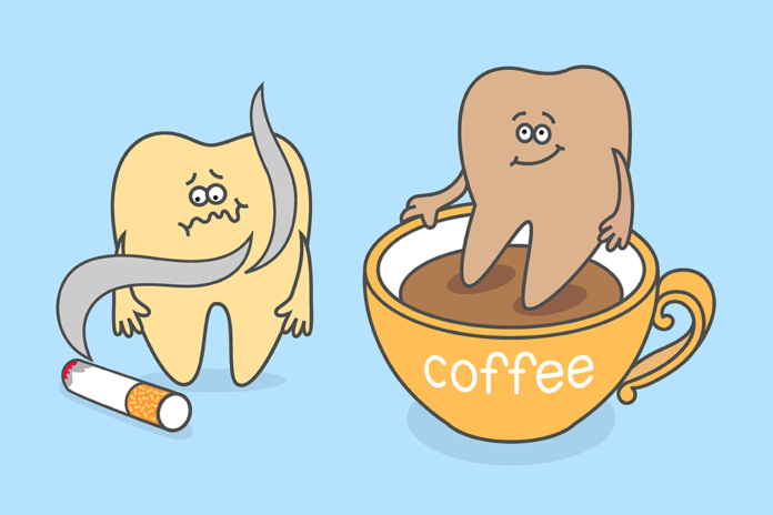
Extrinsic stain ‒ Extrinsic stains are found on the external surfaces of the tooth and can be removed manually, whether with toothbrushing, scaling, or polishing. This category of stain is created by exogenous stain, an external stain most often induced from poor dental hygiene, beverages such as coffee and tea, and tobacco habits.1,2
Unfortunately, some extrinsic stains can also be intrinsic as well. For example, smoking tobacco creates both forms of stain as the nicotine is slowly absorbed into the tubules of the enamel, thus creating a discoloration from within as well as leaving an extrinsic stain on the surface.
Extrinsic stains are also classified as metallic and nonmetallic. Nonmetallic are those absorbed onto the tooth’s deposits, such as plaque.1 They generally are comprised of tobacco, beverages, dietary components, and mouth rinses such as chlorhexidine.
A form of bacteria within the nonmetallic classification is called chromogenic bacteria (color-producing bacteria) ‒ also responsible for staining. Chromogenic bacteria staining is commonly seen on the cervical regions of the tooth following the contour of the enamel, presenting in various colors. Children are more likely to exhibit this form of staining; however, it has been discovered in adults.1,3 Studies suggest that in children with poor oral hygiene, the staining presents as orange/green and black/brown in those with good oral hygiene habits.1 Black stain studies confirm that it is a form of bacterial plaque and mainly consists of black insoluble ferric compound, probably ferric sulfide.3
The betel plant is a popularly used plant in Indonesia for both medical and dental purposes. The betel leaf is commonly chewed to treat halitosis, thrush, and dental pain, among other medical purposes.5 Due to the tannins found in this plant, a red-black hue is deposited on the enamel.
Metallic stains are created from exposure to metallic salts, such as the black stains from iron supplements. Mouth rinse is also subject to metallic staining, dependent upon the ingredients. Rinses that contain stannous fluorides often create a golden-brown stain unless the stannous ion is stabilized with zinc phosphate technology that inhibits oxidation (preventing staining).
Yet another form of extrinsic stain comes from the ever-popular sport of swimming. Swimmer’s calculus is the term for the staining that competitive swimmers often exhibit from the frequent exposure to chlorine. Chlorine is used to eliminate or kill harmful microbes such as viruses that may exist. The chemical element elevates the pH levels of the water above the pH that is found in saliva. This causes the salivary proteins to break down faster, creating the perfect environment for organic matter to deposit on the teeth and causing stain.4
The Role of the Dental Professional
It is our role as dental professionals to help determine the source of the stain and offer ways to treat or control it. Some intrinsic stain may not respond to whitening agents successfully, and the dental professional will need to be readily prepared to discuss cosmetic treatment such as crowns and veneers.
This opens up a perfect opportunity to encourage routine appointments to control plaque, calculus, and stain, which will, in turn, lead to a healthier oral cavity. Offering alternative products that may decrease stains, suggesting whitening agents, and providing education on proper oral hygiene will help assist patients with their staining frustrations.
| Color of Stain | Source |
| Brown stain | -Due to tannin
-Often found in coffee, tea, and chromogenic bacteria -Chlorhexidine rinse |
| Black stain | -Coal tar combustion products such as tobacco
-Exposure to metals such as iron -Chromogenic bacteria |
| Orange stain | -Chromogenic bacteria found in children |
| Green stain | -Chromogenic bacteria in children
-Decomposed hemoglobin -Copper salts in mouth rinse |
| Golden brown stain | -Exposure to stannous fluoride |
| Red-black stain | -Use of betel leaves and nuts |
Before you leave, check out the Today’s RDH self-study CE courses. All courses are peer-reviewed and non-sponsored to focus solely on high-quality education. Click here now.
Listen to the Today’s RDH Dental Hygiene Podcast Below:
References
- Prathap, S., Hosdurga, R., Boolor, A., Rao, S. (2013). Extrinsic Stains and Management: A New Insight. J. Acad. Indus. Res. 2013; 1(8): 45-42. Retrieved from http://jairjp.com/JANUARY%202013/02%20SRUTHY%20PRATHAP.pdf
- Sahi, A. (2018, August 23). Tooth Polishing Procedure. News Medical Life Sciences. Retrieved from https://www.news-medical.net/health/Tooth-Polishing-Procedure.aspx
- Prathap, S., Prathap, M.S. Occurrence of Black Chromogenic Stains and its Association with Oral Hygiene of Patients. Ann Med Health Sci Res. 2018; 8: 18-21. Retrieved from https://www.amhsr.org/articles/occurrence-of-black-chromogenic-stains-and-its-association-with-oral-hygiene-of-patients-4355.html
- Howley, E. (2021, February 5). How Can I Keep My Teeth White as a Swimmer? U.S. Masters Swimming. Retrieved from: www.usms.org/fitness-and-training/articles-and-videos/articles/how-can-i-keep-my-teeth-white-as-a-swimmer
- Hoppy, D., Noerdin, A., Irawan, B., Soufyan, A. Effect of Betel Leaf Extract Gel on Color Change in the Dental Enamel. Journal of Physics: Conference Series. 2018; 1073(3): 032028. Retrieved from https://iopscience.iop.org/article/10.1088/1742-6596/1073/3/032028/pdf
- Jacobsen, P.E., Henriksen, T.B., Haubek, D., Østergaard, J.R. Developmental Enamel Defects in Children Prenatally Exposed to Anti-epileptic Drugs.” PloS one. 2013; 8(3): e58213. doi:10.1371/journal.pone.0058213. Retrieved from https://journals.plos.org/plosone/article?id=10.1371/journal.pone.0058213


