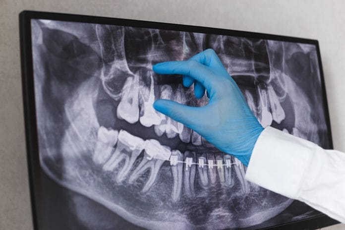
The Archives of Clinical Skiagraphy was the first radiology scientific journal. First published in May 1896, just six months after the discovery of x-rays by Wilhelm Roentgen, Sydney Rowland wrote in the first editorial: “The object of this publication is to put on record in permanent form some of the most striking applications of the New Photography to the needs of Medicine and Surgery.”5
And, he was right. Radiology has advanced medicine and dentistry in ways that Wilhelm Roentgen, Marie Curie, and Pierre Curie could have never imagined.
If you have never taken the time to look through archived journals such as this one, I recommend you add it to your to-do list. It is absolutely fascinating how much we have advanced in this area of science. The history of radiology is downright amazing and brimming with fascinating individuals.
Although I could go on for days about the history of radiology, this is meant to be an update, not a review. Technology is advancing so rapidly it is often hard to keep up. When I was in hygiene school, we still used films and even had a unit in radiology that required us to develop radiographs by hand-dipping.
Times have certainly changed since then with digital radiographs. These advancements have also brought change to many of the guidelines for dental radiographs.
Radiographs During Pregnancy
In the past, before E and F speed film and digital radiographs, taking radiographs during pregnancy was contraindicated. Because of the tremendous reduction in radiation when using faster speed film and digital radiographs, there are no longer any contraindications. According to the ADA and the ACOG, “X-rays with shielding of the abdomen and thyroid is safe during pregnancy.”1
Of course, informed consent from the patient is ideal. The clinician should explain the importance and safety of taking dental radiographs. The clinician should also follow the ALARA principle. Pregnant women are at an increased risk for dental caries, and this should be taken into consideration when deciding if radiographs are necessary.2
Some studies indicate that lead shielding is not necessary for dental radiographs for pregnant patients. A study published in 2016 measured the dose of radiation to the fetus and breasts with lead shielding and without lead shielding. The study found the dose with or without the lead shield was far below the level associated with harm to the fetus. The authors did add that this was technique specific.6
Every clinician should follow, of course, the ADA and ACOG’s guidelines and use a lead shield. However, studies like this may help clinicians feel more comfortable when taking radiographs on pregnant patients. The knowledge that the radiation dose is well below a dose that could cause harm to the fetus may also be reassuring for the patient.
Lead Shield Use
Lead shielding of patents is a practice that hygienists have followed for many years. Some feel it is reassuring to the patient and for their own peace of mind. However, are lead shields necessary to protect patients from radiation?
According to the Health Physics Society, “Use of the lead apron to protect the patient undergoing dental radiographic examination was recommended some 50 years ago when equipment was crude. This was because x-ray beams were not restricted to the area of clinical interest, beams were not filtered, and x-ray film was slower, causing radiation exposures 10 to 100 times higher than received today. With the current technology reducing radiation exposure significantly and the beam limited only to the area of interest, there is little or no measurable difference in whole-body dose whether a lead apron is used or not. The lead apron is no longer regarded as essential, although some consider it a prudent practice, especially for pregnant and potentially pregnant females.”7
This stance is supported by many scientific studies. One study evaluated the absorbed dose from a panoramic radiograph with a lead shield and without a lead shield. The study found no statistically significant difference in the amount of absorbed dose with or without the use of a lead shield.3
Additionally, at a board of directors meeting in April 2019, the American Association of Physicists in Medicine recommended the discontinued use of lead shielding. In the position statement released, the organization stated, “Patient shielding is ineffective in reducing internal scatter. In medical x-ray imaging, the main source of radiation dose to internal organs that are outside the imaging field of view is x-rays that scatter inside the body. However, surface shielding covering these organs has no impact on this scatter.”4
Many of these scientists believe we may be perpetuating an ongoing fear and misunderstanding of radiation leading to patients declining radiographs. The statement “this is the way we have always done it” or “what is the harm? It makes patients more comfortable” is often muttered by dental professionals not wanting to change.
Yet many of these same clinicians get frustrated when patients refuse their routine radiographs. There has been a fear instilled in many people about radiation exposure in dentistry. If clinicians are against implementing change backed by science, how can we expect patients to change their way of thinking?
To make the topic even more complicated, current guidelines for lead shield use is governed by each individual state. Some states recommend or require use, while others do not. This is also a problem and sends a mixed message to patients. This can be confusing for hygienists that communicate across state lines on social media as well. Although currently there doesn’t seem to be a clear consensus, don’t be surprised if in the very near future, dentistry retires the use of the lead shield altogether.
Frequency of Radiographs
It seems as if some offices and clinicians view the frequency of radiographs as a mandated annual event, nothing could be further from the truth. The ADA collaborated with the FDA to develop specific guidelines that require risk assessment when determining frequency of radiographs. These guidelines consider age, dental development stage, new patients, recall patients, and caries/periodontal risk. The chart is available on the ADA’s website here.
For example, children and adolescents who are recall patients with increased caries risk should get radiographs every six to 12 months. Recall adult patients with increased caries risk should get radiographs every six to 18 months.
In contrast, low caries risk children with primary and mixed dentitions should have radiographs taken every 12 to 24 months. Low caries risk adolescents with permanent dentition should have radiographs every 18 to 36 months, and low caries risk adults should have radiographs every 24 to 36 months.
As you can see, it is meant to be decided on an individual basis what the frequency should be for each patient.
Universal frequencies, like once a year, may not be adequate for some patients and maybe too much for others. Hygiene school prepared us to use critical thinking skills, and this is a perfect time to put them in use. Of course, as hygienists, we always have the dentist to help in making these decisions, since ultimately, he/she will be the prescriber of the radiographs.
Conclusion
In the time it takes for this article to be published, there could be more changes. That is how fast the science of radiology is progressing. It is difficult to stay in the know with such a quickly progressing area of science and technology. I hope this article will help clear up some of the confusion about current radiology practices in dentistry. Taking regular CE courses on some of these topics that are quickly advancing may be helpful for clinicians that remain uncertain of proper radiation safety and protocol.
Now Listen to the Today’s RDH Dental Hygiene Podcast Below:
References
- Oral Health Topics. Xrays/Radiographs. American Dental Association. Retrieved from https://www.ada.org/en/member-center/oral-health-topics/x-rays
- Pregnancy and Oral Health. Centers for Disease Control and Prevention. Retrieved from https://www.cdc.gov/oralhealth/publications/features/pregnancy-and-oral-health.html
- Rottke, D., Grossekettler, L., Sawada, K., Poxleitner, P., Schulze, D. Influence of Lead Apron Shielding on Absorbed Doses from Panoramic Radiography. Dentomaxillofac Radiol. 2013; 42(10): 20130302. DOI:10.1259/dmfr.20130302 Retrieved from https://www.ncbi.nlm.nih.gov/pmc/articles/PMC3852527/
- AAPM Position Statement on the Use of Patient Gonadal and Fetal Shielding. American Association of Physicists in Medicine. Retrieved from https://www.aapm.org/org/policies/details.asp?id=468&type=PP
- Rowland S. Archives of clinical skiagraphy. 1896. Br J Radiol. 1995;68(805):H1–H20. doi:10.1259/0007-1285-68-805-H1. Retrieved from https://pubmed.ncbi.nlm.nih.gov/7881874-archives-of-clinical-skiagraphy-1896/
- Kelaranta, A., Ekholm, M., Toroi, P., Kortesniemi, M. Radiation Exposure to Fetus and Breasts from Dental X-ray Examinations: Effect of Lead Shields. Dentomaxillofac Radiol. 2016; 45(1): 20150095. DOI:10.1259/dmfr.20150095 Retrieved from https://www.ncbi.nlm.nih.gov/pmc/articles/PMC5083886/
- Health Physics Society Specialists in Radiation Protection. Dental Patient Issues. Retrieved from http://hps.org/publicinformation/ate/faqs/dentalpatientissuesqa.html

