Obstructive sleep apnea (OSA) is characterized by recurrent episodes of partial or complete upper airway obstruction, often caused by the collapse of the airway due to anatomical and functional factors. People most predisposed to developing OSA are overweight or obese adult males with mandibular retrusion, micrognathia, or maxillofacial alterations. It is estimated that 936 million adults worldwide are affected by OSA.1
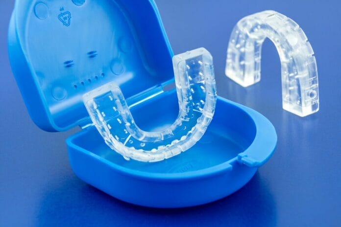
The gold standard treatment for OSA is continuous positive airway pressure (CPAP). However, compliance with the long-term use of CPAP is poor. There are alternative options for patients who cannot tolerate using a CPAP. One such therapeutic alternative that can be acquired from the dentist is a mandibular advancement device (MAD).1
MADs consist of two acrylic splints that interface with each other. The mechanism of action differs depending on the appliance design. Nonetheless, the goal of the MAD is to generate mandibular protrusion to prevent airway collapse. The effectiveness of MADs is comparable to that of CPAP in patients with mild or moderate OSA, but inferior to CPAP in patients with severe OSA. Compliance with MADs is superior to CPAP, which allows it to remain an option for patients with severe OSA who cannot tolerate CPAP.1
Unfortunately, there are contraindications for using MADs, including severe temporomandibular joint problems, active periodontal disease, grade 2 or 3 tooth mobility, an insufficient number of healthy teeth to retain the device, or poor jaw movement.1
Although moderate to severe periodontal disease is a well-established contraindication for MADs, there is limited research on their effects on the periodontium. While previous studies have shown that bone loss increases tooth stress and movement, most have focused only on a single tooth and have not included MADs. Consequently, there is a gap in knowledge regarding the effects of MADs on the entire periodontium in patients with a healthy periodontium and those with early-stage periodontitis.1
A study aimed to fill that gap by testing four commercial MADs under two conditions, healthy bone and early-stage periodontitis (15% bone loss), to evaluate how reduced bone support changes the impact of MADs on teeth and periodontal ligaments.1
The Study
The study used computer modeling called finite element analysis (FEA) to predict the effects of MADs on the periodontium of healthy and early-stage periodontitis. A 3D model was created based on an anatomical model obtained from a high-resolution computed tomography (CT) scan of a healthy 29-year-old female patient. The hard tissues were modeled using Mimics software, and since soft tissues aren’t easily identified in CT scans, they were added using Rhinoceros computer-aided design (CAD) software. The PDL was designed as a thin 0.3 mm layer between the tooth and the alveolar socket of the 3D model. Physical prototypes of the MADs were scanned using a Konica Minolta laser scanner.1
To simulate early-stage periodontitis, bone and PDL were removed. The amount removed was determined based on the root length, using the metric of the distance from the cementoenamel junction (CEJ) to the apex of the tooth to create a 15% loss of supporting tissue.1
The final model for FEA included the teeth of both arches, PDL for each tooth, and one of four MADs. The maxillary and mandibular bones were excluded from the FEA because it was believed that, due to the stiffness and rigidity of the bone compared to the PDL, there would be little effect on the study’s results. The authors also verified that including the bone did not change the stress results on the PDL, which justified its exclusion from the model.1
Four MADs with different mechanisms of action were included in this study. The four mechanisms evaluated were MADS with telescopic arms, buccal inclined extensions, adjustable straps, and anterior connecting rods. Each design has different designated connection points and mechanisms for mandibular protrusion.1
Since these appliances are custom-fitted for each patient, the anatomical models were scanned using a laser scanner and converted into a 3D digital model of the MAD splint. After the splint was created, the mechanism specific to each MAD was added (rods, arms, straps, etc.) The MADs were then adjusted to fit using digital modeling techniques.1
For the simulation, all materials used for the MADs replication were assigned values for stiffness and stretch. The materials were assumed to have linear elasticity in all directions. Although the four MADs utilized in this study are made from different materials, the study used a standard hard acrylic material for a fair comparison.1
The PDL was not modeled as a simplistic linear elastic material, unlike the other structures, due to its nonlinear nature. Instead, a hyperelastic model was used to simulate its behavior under stress, as it does not behave like a typical solid material. This was based on a strain energy density function mathematical model to represent the complex mechanical properties of the tissue.1 The mathematical model used was a second-order Ogden model, which describes the nonlinear stress-strain behavior of materials such as rubber, polymers, and soft tissue.1,2 This model allows researchers to assess the strain imposed by specific stresses on soft tissues, in this case, the replicated PDL.1
The PDLs were given fixed support to mimic the maxillary and mandibular bone. This measure provided support and prevented rigid body motion of the model during the static structure analysis, which determined how the structures would behave under constant, non-changing load. While this constraint prevented the model from moving as a whole, it was necessary to accurately measure the stress and deformation that were applied to the teeth and PDLs.1
The teeth and MAD appliance were given a bonded contact to prevent slipping or separation. The authors noted that this was a reasonable assumption because the device’s retention force, due to its interproximal fit on the teeth, would be greater than the applied load. While this bonded condition simplified the simulation and allowed for faster convergence, the model also accounted for the PDLs providing the necessary support for the teeth to withstand the forces of clenching, bruxing, and the tight fit of MADs over the teeth.1
To simulate mandibular advancement, a forward mandibular jaw movement of 9.5 mm, the typical movement for treating OSA, was modeled. The force used to simulate mandibular advancement was 5.59 N (newtons). Static, equal, and opposite forces were applied at the connection points between the splints, allowing for consistent comparison. Eight total simulation models were created, and each MAD was used in both a healthy model and a model based on early-stage periodontitis, using identical setups for an impartial comparison between the MADs.1
The Results
The results showed that MADs influence both stress and tooth movement:1
-
- MADs using adjustable straps exhibited stress on mandibular molars and the maxillary anterior teeth and produced the most tooth movement, reaching a displacement value of 0.0019 mm.
- MADs using anterior connecting rods exhibited stress and movement, mainly in the anterior teeth and premolars.
- MADs using buccal inclined extensions exhibited lower and more evenly distributed stress, mainly affecting maxillary molars and mandibular lateral incisors and canines.
- MADs using telescopic arms exhibited the lowest displacement despite targeting maxillary premolars and mandibular anterior teeth. This design was the least aggressive of all tested MADs (displacement value of 0.0011 mm).
The study found a slight increase in stress and displacement in the early-stage periodontitis models compared to the healthy models, with a more significant increase in the maxillary teeth. The overall stress was higher in the pathological models due to tissue loss. The maxillary PDLs showed a greater increase in stress because the average tissue loss was more significant in the maxilla (20% loss versus 17% loss in the mandible).1
Specifically, the percentage increase in PDL stress in the maxilla for each MAD was as follows:1
-
- Adjustable straps: 21%
- Buccal inclined extensions: 17%
- Anterior connecting rods: 16%
- Telescopic arms: 7%
Mandibular PDL stress was significantly less than maxillary PDL stress. The most significant stress on the mandible was observed with MADs using anterior connecting rods and telescopic arms, both of which showed a 7% increase. In contrast, the other mechanisms produced no significant changes, with an increase ranging from 0% to -1%. The stress patterns were similar in the mandible between healthy and early periodontitis models.1
Discussion
This study aimed to expand the current knowledge on the effects of MAD designs on the periodontium of healthy patients and those with early-stage periodontitis. The focus was on how MADs affect teeth and the PDL. Unlike previous studies on a single tooth, this study used full arch models.1
A prolonged increase in mechanical load on teeth can cause acute inflammation that may become chronic. This process can inhibit the recruitment of cells necessary for bone remodeling, which can affect tooth movement. The stress exerted on the teeth and PDLs by MADs is therefore a significant consideration, as the study’s findings suggest that increased stress on PDLs may worsen pre-existing periodontal conditions and necessitate long-term oral health monitoring.1
The findings are supported by a previous study that found MADs with a more anterior protrusion mechanism produced more pronounced occlusal changes than those designed with a bilateral thrust. Although the study showed that changes in tooth position were minimal, the authors noted that over time, these small shifts could contribute to alterations in a patient’s occlusion, potentially having functional and aesthetic implications.1
This study has multiple limitations:1
-
- Material uniformity: All MADs were modeled with the same material, which might not reflect actual differences.
- Single advancement step: The simulation used a single fixed mandibular advancement of 9.5 mm, while in a real-world setting, this process is typically done gradually.
- Model symmetry: The model assumes perfect symmetry between the left and right sides, which may not reflect reality.
- Bonded contact: A bonded connection between the teeth and the MAD was utilized, which is unrealistic.
- Uniform bone loss: Bone loss was modeled evenly across all teeth, rather than reflecting individual variations.
- Exclusion of soft tissue: Soft tissues were not included, which eliminated probing depth and clinical attachment loss as potential metrics to assess the effects.
Conclusion
Although compliance with MADs is reportedly better than that of CPAP for OSA, there are contraindications for use. Previous studies had focused mainly on single-tooth models and had not correlated the effects of MADs with periodontitis. This study fills that gap by providing a comprehensive comparison of different MADs on the entire periodontium of patients with and without early-stage periodontitis. However, the authors note that additional studies are needed to better investigate more severe stages of the disease, account for inter-tooth variability in bone loss, and model the gradual advancement of the mandible.1
The results have clinical significance for occlusion, tooth position, and the health of the PDL. MADs with lateral mechanisms, such as telescopic arms, distribute less stress more evenly and may be better suited for patients with early-stage periodontitis. Meanwhile, MADs using buccal inclined extensions and anterior connecting rods may worsen periodontal conditions. Therefore, it is essential to monitor patients using MADs throughout treatment, even in those with a healthy periodontium.1
Before you leave, check out the Today’s RDH self-study CE courses. All courses are peer-reviewed and non-sponsored to focus solely on high-quality education. Click here now.
Listen to the Today’s RDH Dental Hygiene Podcast Below:
References
- Caragiuli, M., Candelari, M., Zalunardo, F., et al. Effects of Oral Appliances for Obstructive Sleep Apnoea in Reduced Periodontium: A Finite Element Analysis. Int Dent J. 2024; 74(6): 1306-1316. https://pmc.ncbi.nlm.nih.gov/articles/PMC11551564/
- Lohr, M.J., Sugerman, G.P., Kakaletsis, S., et al. An Introduction to the Ogden Model in Biomechanics: Benefits, Implementation Tools and Limitations. Philos Trans A Math Phys Eng Sci. 2022; 380(2234): 20210365. https://pmc.ncbi.nlm.nih.gov/articles/PMC9784101/


