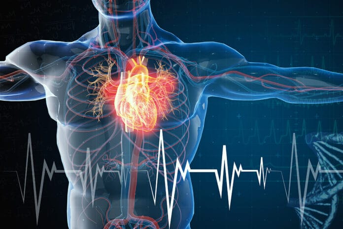
Every 37 seconds, cardiovascular disease (CVD) kills someone in the United States – more than all cancers combined.1 Even though death rates for coronary heart disease have dropped by two-thirds during the past 40 years, CVD still accounts for 35% of all American deaths per year and is clinically diagnosed in 80 million Americans.1
While the absolute number of deaths due to CVD has decreased among males, it has increased among females.1 The number one cause of death and the number one cause of disability among both men and women in this country is CVD.2
The most common manifestations of CVD are connected directly to the heart, such as chest pain from ischemia and edema caused by the heart’s reduced capacity to pump blood. Other dominant symptoms of heart disease include labored breathing, palpitations, hypotension, and syncope.3 Most healthcare practitioners are aware of these familiar signs. There are other, more unusual signs of CVD that we should be aware of as well.
The following signs may indicate that your patient has heart disease, which might be undiagnosed and untreated. By being cognizant, bringing these indicators to the patient’s awareness, and referring your patient to a cardiologist, you can help improve your patient’s quality of life and longevity. Twenty-five percent of all deaths due to CVD are sudden, so your keen eye for recognizing possible subclinical stages of atherosclerosis could make the difference between life and death.4
Keen Eye of Dental Hygienists Can Spot Unusual Signs
Imagine being able to help your patient adopt a healthier lifestyle and prevent the progression of heart disease because you are an astute, whole health dental hygienist. This is one way we save lives in the dental office.
1) Frank‘s sign (diagonal earlobe crease)
Numerous studies point to a correlation between a diagonal crease in the earlobe with a higher frequency of cardiac events.5 There is still debate among experts about the reliability of Frank’s sign as an indicator of CVD; however, most agree it could be an important signal. The crease extends at least one-third the distance from the tragus to the auricle.
This sign is more common among the elderly but also observed in younger people. It is thought to be due to age-related and microvascular disease associated with weakening dermal and vascular elastic fibers. A diagonal ear crease is easily detectable (unless your patient is wearing earrings) and may facilitate prompt evaluation and early diagnosis of coronary atherosclerosis, especially in the presence of other concurrent risk factors.
2) Forehead wrinkles
According to research presented at the European Society of Cardiology Congress 2018, people who have many deep forehead wrinkles that are more than expected for their age may be at a higher risk of dying from CVD.6 The researchers followed 3,200 working adults for 20 years.
Wrinkle scores based on the number and depth of the wrinkles were assigned. The investigators discovered that people with a wrinkle score of one had a slightly higher risk of dying of CVD than people who had no wrinkles. People who had wrinkle scores of two and three had almost 10 times the risk of dying of CVD compared to people who had wrinkle scores of zero. They concluded that the higher the wrinkle score, the higher the risk of cardiovascular mortality.
The investigators hypothesized that horizontal forehead wrinkles could be related to plaque accumulation and atherosclerosis. Arteries in the forehead are small and susceptible to plaque accumulation, and wrinkles are an early sign of vessel senescence.
Additionally, collagen protein changes and oxidative stress are both factors in wrinkle development as well as atherosclerosis. According to the authors of the study, forehead furrows are not a better method of measuring cardiovascular risk than existing methods, but they could raise a red flag and allow for earlier discovery of a potentially life-threatening problem.
On a side note, crow’s feet, the wrinkles that are found at the outside corners of the eyes, are not connected to an increased risk of CVD.7
3) Xanthelasma (cholesterol build-up on eyelids) and xanthomas (cholesterol build-up on other areas of the skin)
Xanthelasma and xanthomas are yellowish growths seen on the eyelids, elbows, knees, and buttocks. They are a sign of hyperlipidemia, which is an excess of fat in the bloodstream.
According to a 2011 study in the British Medical Journal, xanthelasmas are associated with dyslipidemia, atherosclerosis, and coronary artery disease.8 The study found additional associations with hypertension, central obesity, and diabetes. The authors state that xanthelasmas predict the risk of CVD and death in the general population, independently of well-known CVD risk factors.
4) Vertex baldness (male pattern baldness)
Vertex baldness, more commonly known as male pattern baldness, originates at the crown of the head. A correlation has been made between this particular pattern of hair loss and an increased risk of CVD.9 This same association has not been identified between frontal baldness and CVD.
According to the authors, the strength of the correlation depends on the degree of vertex hair loss. A person with more significant vertex hair loss has a higher risk of CVD than someone with less vertex hair loss. An association has been found among younger men who exhibit this pattern of baldness as well.
5) Digital clubbing (clubbed nails)
Digital clubbing is characterized by bulbous enlargement of the ends of the fingers and/or toes. According to a study by Sarkar et al., it is the result of hypertrophy of soft-tissue components of the pulp and hyperplasia of the fibrovascular tissue at the nail base.10
The authors state that clubbed nails may be a predictor of a variety of underlying diseases such as lung cancer, cirrhosis of the liver, cyanotic congenital heart disease, aortic aneurysm, and congenital or acquired CVD. Sarkar et al. reported that digital clubbing progresses as oxygen levels decrease.10
6) Arcus senilis (halo around the iris)
Arcus senilis is a blue, gray, or white arc that is visible above and below the outer part of the cornea ‒ the clear, domelike covering over the front of the eye. The arc may eventually form a complete ring around the iris, or colored portion, of the eye.
This halo is common in older adults and is caused by lipid deposits deep in the edge of the cornea. Early studies found that arcus senilis was considered a strong predictor of coronary heart disease and CVD mortality. Yet, later studies found it was only a predictor of CVD in relation to advancing age.11 Arcus senilis does not affect vision and does not require treatment; however, awareness of this sign may be useful in early intervention in CVD.
7) Cyanosis (blue lips)
Cyanosis is the medical term for discoloration of the lips, skin, tongue, or other mucous membranes. It is caused when the body does not receive enough oxygenated blood. In Caucasian people, cyanosis causes tissues to turn blue. In African American people, cyanosis may cause the tissues to turn gray or whitish.
Regardless of the exact appearance of cyanosis, it is an indication of poor blood flow.12 Cyanotic changes to skin or lip color indicate a blockage in a blood vessel. Without treatment, a lack of blood and oxygen supply can result in the death of underlying tissues.13
8) Edema (swelling of lower legs, ankles, and feet)
Many diseases of the cardiovascular system can result in edema or fluid build-up in the lower extremities.14 This fluid build-up is a result of chronic venous insufficiency or reduced ability of the veins to return blood to the heart. Edema, or swelling of the lower limbs, is reported to be highly predictive of CVD.15
In warmer months, when patients may present for dental appointments in shorts, skirts, and sandals, we have an opportunity to observe these lower extremities for signs of edema.
9) Livedo reticularis (blue or purple net-like pattern on the skin)
This net-like pattern on the skin may be reminiscent to dental professionals of the reticular form of lichen planus. While reticular lichen planus appears as white lacey patches on red mucosal tissues, livedo reticularis appears as a blue or purple lacey or net-like pattern on the skin, most commonly on the legs.16
It is seen in people who have a serious condition called cholesterol-embolization syndrome (CES), a manifestation of atherosclerosis.17 Livedo reticularis is also seen in people who have an autoimmune disease called antiphospholipid antibody syndrome.16 In CES, small arteries become blocked, which may lead to damaged tissues and organs, and may result in CVD and death.18 If you notice this type of lacey pattern on your patient’s skin, and the patient does not complain of feeling cold, a medical referral may be in order.
10) Poor oral health
Periodontal disease, dental caries disease, and tooth loss have been linked to a higher risk of CVD.19 For years, experts believed a possible explanation for the link between poor oral health and heart disease could be that bacteria from the mouth travel to arteries in the body, causing blood vessel damage and inflammation, which can contribute to heart disease.
In 2016, a landmark study published by Bale, Doneen, and Vigarust demonstrated level 1 scientific proof that this is the case.20 The researchers found that certain high-risk oral pathogens cause atherosclerosis. With the availability, ease, and affordability of bacterial DNA testing, we can now determine if our patients harbor any pathogenic bacteria in their oral microbiome, and we can target treatment to reduce and even eliminate these pathogens. In this way, we can positively affect the course of our patient’s cardiovascular disease risk.
As dentistry moves towards practicing more holistic health care, we must observe systemic signs and symptoms of disease. By being astutely aware of these 10 curious signs of cardiovascular disease, dental professionals increase our ability to recognize and mitigate patients’ risk for CVD and death.
As I always say, “We’re not just cleaning teeth, we’re saving lives.”
Now Check Out the Peer-Reviewed, Self-Study CE Courses from Today’s RDH!
Listen to the Today’s RDH Dental Hygiene Podcast Below:
References
- Wall, H.K., Ritchey, M.D., Gillespie, C., et al. Vital Signs: Prevalence of Key Cardiovascular Disease Risk Factors for Million Hearts 2022 – United States, 2011-2016. MMWR Morb Mortal Wkly Rep. 2018; 67(35): 983-991. Published 2018 Sep 7. doi:10.15585/mmwr.mm6735a4.
- Woodward, Cardiovascular Disease and the Female Disadvantage. Int J Environ Res Public Health. 2019; 16(7): 1165. Published 2019 Apr 1. doi:10.3390/ijerph16071165.
- Flora, G.D., Nayak, M. A Brief Review of Cardiovascular Diseases, Associated Risk Factors and Current Treatment Regimes. Curr Pharm Des. 2019; 25(38): 4063-4084. doi: 10.2174/1381612825666190925163827. PMID: 31553287.
- McElwee, S.K., Velasco, A., Doppalapudi, H. Mechanisms of sudden cardiac death. J Nucl Cardiol. 2016 Dec; 23(6): 1368-1379. doi: 10.1007/s12350-016-0600-6. Epub 2016 Jul 25. PMID: 27457531.
- Baboujian, A., Bezwada, P., Ayala-Rodriguez, Diagonal Earlobe Crease, a Marker of Coronary Artery Disease: A Case Report on Frank’s Sign. Cureus. 2019; 11(3): e4219. Published 2019 Mar 11. doi:10.7759/cureus.4219.
- The abstract “Forehead wrinkles and risk of all-cause and cardiovascular mortality over 20-year follow-up in working population: VISAT study” was presented duringPoster session 2: Risk assessmenton Sunday 26 August, 2018.
- Christoffersen, M., Tybjaerg-Hansen, A. Visible aging signs as risk markers for ischemic heart disease: Epidemiology, pathogenesis and clinical implications. Ageing Research Reviews. 2016; 25: 24-41.
- Christoffersen, M., Frikke-Schmidt, R., Schnohr, P., et al. Xanthelasmata, arcus corneae, and ischaemic vascular disease and death in general population: prospective cohort study. BMJ. 2011; 343: d5497.
- Pechlivanis, S., Heilmann-Heimbach, S., Erbel, R., et al. Male-pattern baldness and incident coronary heart disease and risk factors in the Heinz Nixdorf Recall Study. PLoS One. 2019; 14(11): Published 2019 Nov 19. doi:10.1371/journal.pone.0225521.
- Sarkar, M., Mahesh, D.M., Madabhavi, Digital clubbing.Lung India. 2012; 29(4): 354-362. doi:10.4103/0970-2113.102824.
- Fernandez, A.B., Keyes, M.J., Pencina, M., et al. Relation of corneal arcus to cardiovascular disease (from the Framingham Heart Study data set). Am J Cardiol. 2009; 103(1): 64-66. doi:10.1016/j.amjcard.2008.08.030.
- Pahal, P., Goyal, A. Central and Peripheral Cyanosis. [Updated 2021 Oct 9]. In: StatPearls [nternet]. Treasure Island (FL): StatPearls Publishing; 2022 Jan. https://www.ncbi.nlm.nih.gov/books/NBK559167/
- Uliasz, A., Lebwohl, Cutaneous manifestations of cardiovascular diseases. Clin Dermatol. 2008 May-Jun; 26(3): 243-54. doi: 10.1016/j.clindermatol.2007.10.014. PMID: 18640521.
- Kataoka, Clinical characteristics of lower-extremity edema in stage A cardiovascular disease status defined by the ACC/AHA 2001 Chronic Heart Failure Guidelines. Clin Cardiol. 2013 Sep; 36(9): 555-9. doi: 10.1002/clc.22159. Epub 2013 Jul 10. PMID: 23843030; PMCID: PMC6649399.
- Prochaska, J.H., Arnold, N., Falcke, A., et al. Chronic venous insufficiency, cardiovascular disease, and mortality: a population study. Eur Heart J. 2021 Oct 21; 42(40): 4157-4165. doi: 10.1093/eurheartj/ehab495. PMID: 34387673.
- Sajjan, V.V., Lunge, S., Swamy, M.B., Pandit, A.M. Livedo reticularis: A review of the literature. Indian Dermatol Online J. 2015; 6(5): 315-321. doi:10.4103/2229-5178.164493.
- Ozkok, A. Cholesterol-embolization syndrome: current perspectives. Vasc Health Risk Manag. 2019; 15: 209-220. Published 2019 Jul 8. doi:10.2147/VHRM.S175150.
- Soltész, P., Szekanecz, Z., Kiss, E., Shoenfeld, Y. Cardiac manifestations in antiphospholipid syndrome. Autoimmun Rev. 2007 Jun; 6(6): 379-86. doi: 10.1016/j.autrev.2007.01.003. Epub 2007 Jan 31. PMID: 17537384.
- Kotronia. E., Brown, H., Papacosta, A.O., et al. Oral health and all-cause, cardiovascular disease, and respiratory mortality in older people in the UK and USA. Sci Rep. 2021; 11(1): 16452. Published 2021 Aug 12. doi:10.1038/s41598-021-95865-z.
- Bale, B.F., Doneen, A.L., Vigerust, D. High-risk periodontal pathogens contribute to the pathogenesis of atherosclerosis. Postgrad Med J. 2017 Apr; 93(1098): 215-220. doi: 10.1136/postgradmedj-2016-134279. Epub 2016 Nov 29. PMID: 27899684; PMCID: PMC5520251.


