A few years ago, our dental office implemented a microscope for the hygiene room. Game changer! I had no idea what a profound impact the instrument would have on dental hygiene treatment.
There is much more awareness of the oral-systemic link these days. When discussing the oral-systemic link, we must discuss the oral microbiome. Biofilm is the collection of various microorganisms adhering and growing on a surface. That is the very simple, generic definition.
In a dental biofilm, bacteria, viruses, fungi, and parasites can be present. Regular disruption of these biofilms is paramount in the prevention of disease. I have been a practicing hygienist for 25 years, and I tell my patients all the time, “Science has proven the mouth is indeed connected to the rest of the body.”
With the microscope, we can visually see for ourselves and show the patients what is in their biofilm.
Using a Microscope
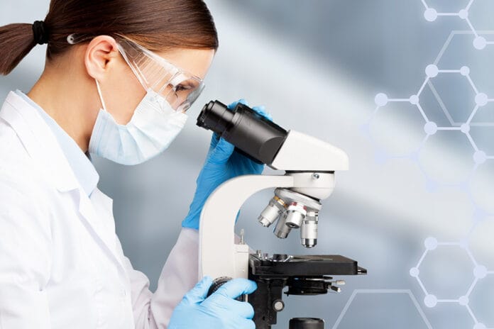
The microscope is a phenomenal, objective tool to determine exactly what is in a patient’s oral biofilm at any given moment. It is a phase-contrast microscope set up in any operatory, although the hygiene rooms are perfect. Optimally, it would have a monitor attached so that both the hygienist and patient can view the samples together while the patient is seated in the chair.
Once the patient is seated, a subgingival plaque sample is collected and placed on a microscope slide. A slide cover is put on top, and then the sample is ready for the microscope. The microscope is connected to a TV monitor so both the patient and the hygienist can view it together.
At this point, the hygienist explains what is happening in the sample. What exactly can be seen on a microscope slide of dental plaque? Various types of bacteria, fungus, parasites, white blood cells, and red blood cells. When reviewing the sample, we first ask the question, is it healthy overall? What does a healthy sample even look like? Is it at risk?
When the patient sees what is actually swimming around in their mouths with their own eyes, they are more far more apt to take action.
Typically, they are fascinated and can’t get enough. Honestly, with some really “good” ones, I can’t get enough either. I want to sit back, grab some popcorn, and watch that amoeba engulf that white blood cell as if I have nothing else to do all day. I want to see those snake-like spirochetes attacking white blood cells, red blood cells, other bacteria … everything. They attack it all!
However, like everyone else, I have more than one patient on the schedule and have to forego the popcorn party and move on.
As soon as the sample has been viewed, this is when the questions begin. “Is that the worst you’ve ever seen?” “How did those get there?” “How do we get rid of those?” “When can we check to make sure they are gone?”
The questions open up a whole world of discussion, starting with the oral effects and leading to systemic effects. If the biofilm is healthy, this is a great tool to verify that what the patient is doing each day is working. If the sample is at risk, the patient is now more likely to take ownership and is more actively engaged in the treatment of their periodontal disease.
Oral-Systemic Link
Dental plaque is a veritable treasure trove of information. When we are discussing the oral-systemic link, we should not overlook the opportunity to evaluate the oral biofilm. The oral and gut microbiome overlap by 45%.1
If certain bacteria such as spirochetes are present in biofilm, bleeding will always be present as these are extremely inflammatory.2 We have all seen those patients who present with very little plaque yet still have heavy bleeding. I am not talking about the patients who only brushed once in the last six months. We all know what that looks like. I am talking about the ones who have what appears to be good oral hygiene, yet each time we see them in the hygiene chair, they start to bleed as soon as you touch the tissue.
We scratch our heads and talk about stress, hormones, nutrition, etc. Of course, these are all important factors. However, a biofilm sample would be a crucial piece of the puzzle when determining why a patient always has gingival bleeding while presenting with very minimal plaque. In cases like this, spirochetes are almost always present.
In this situation, it is the biofilm’s quality and not the quantity that is the problem. We would never have known this without the microscope. Furthermore, certain oral spirochetes are inflammatory to the cardiovascular system and cross the blood-brain barrier.3-5 These would increase the risk of systemic diseases such as cardiovascular disease and neurodegenerative diseases. Let’s not forget cardiovascular disease has been the number one killer of Americans for quite some time now.6
All of this information would be reviewed with the patient, and then instructions are given on how to cultivate a healthier oral microbiome. When purchasing a microscope, some come with great training. Some do not. Training would include the mechanics of how to use the microscope. It would also involve education on the science of what is seen with the microscope and even how to implement it in a hygiene program. If the microscope does not come with training, there are hygienists and dentists who specialize in training offices about phase contrast microscopes.
Typically, conversations about the mouth involve mainly bacteria, but there is so much more going on in a biofilm. As previously stated, with the microscope, other things can be detected, including parasites, fungi, and white blood cells. All of these are diagnostic and would not be seen without the use of the microscope.
When parasites and/or fungi are present, this is another clue why the tissue has such inflammation. The biofilm must be returned to a healthy state, and only then is tissue allowed to heal.
A Full Hygiene Schedule
Oral biofilm evaluation builds value into the hygiene appointment. The use of the microscope leads to a full hygiene schedule. Almost all my patients greatly appreciate this technology. They are leaning in, and they are engaged! They want to know how to transform their unhealthy biofilm to a healthy, protective biofilm.
Guess what? They want to know when they can schedule again to get their new and improved biofilm checked. That’s right. The hygiene schedule becomes full of patients who are actively involved with their treatment. Actively involved equals showing up and following through with treatment.
The ability to see what is in the biofilm at every visit is pivotal in periodontal treatment. If we never know there are pathogenic bacteria, parasites, or fungus present, how can we achieve a healthy mouth? With the microscope’s use, the patient and hygienist can see when unhealthy situations are present and make necessary changes before periodontal damage is done. Periodontal outcomes become greatly improved and far more predictable. This is beneficial to the patient for improved oral health and the prevention of systemic diseases as well. We can all agree this is a win-win for everybody.
Phase-contrast microscopic evaluation of the biofilm is invaluable in hygiene recall visits because it is fast, is a great barometer of the success of the patient’s daily oral hygiene routine, builds value into each hygiene visit, and provides a crucial piece of the puzzle to oral wellness. The oral microbiome is an important factor in overall health. After all, a healthy mouth is a healthy body.
Before you leave, check out the Today’s RDH self-study CE courses. All courses are peer-reviewed and non-sponsored to focus solely on high-quality education. Click here now.
Listen to the Today’s RDH Dental Hygiene Podcast Below:
References
- Segata, N., Haake, S.K., Mannon, P., et al. Composition of the Adult Digestive Tract Bacterial Microbiome Based on Seven Mouth Surfaces, Tonsils, Throat and Stool Samples. Genome Biology. 2012; 13(6): R42. doi:10.1186/gb-2012-13-6-r42. Retrieved from https://pubmed.ncbi.nlm.nih.gov/22698087/.
- Visser, M.B., Ellen, R.P., New Insights into the Emerging Role of Oral Spirochaetes in Periodontal Disease. Clinical Microbiology and Infection. 2011; 17(4): 502–512. doi:10.1111/j.1469-0691.2011.03460.x. Retrieved from https://pubmed.ncbi.nlm.nih.gov/21414084/.
- Shi, Q., Zhang, B., Huo, N., Cai, C., Liu, H., Xu, J., Association Between Myocardial Infarction and Periodontitis: A Meta-Analysis of Case-Control Studies. Frontiers in Physiology. 2016; (7): 519. doi:10.3389/fphys.2016.00519. Retrieved from https://www.ncbi.nlm.nih.gov/pmc/articles/PMC5095113/.
- Bale, B.F., Doneen, A.L., Vigerust, D.J., High-Risk Periodontal Pathogen Contribute to the Pathogenesis of Atherosclerosis. Postgraduate Medical Journal. 2017; 93: 215-220. https://pmj.bmj.com/content/93/1098/215.
- Rajeev, R., Dhar, G., Sahu, S., Nayak, N., Mishra, M., Periodontal Disease and Neurodegeneration: The Possible Pathway and Contribution from Periodontal Infections. Journal of Clinical and Diagnostic Research. 2018; 12. doi:10.7860/JCDR/2018/29110.11045. Retrieved from https://www.researchgate.net/publication/322676482_Periodontal_Disease_and_Neurodegeneration_The_Possible_Pathway_and_Contribution_from_Periodontal_Infections.
- Heron, M., Deaths: Leading causes for 2017 pdf icon[PDF – 3 M]. National Vital Statistics Reports. 68(6). Retrieved from https://www.cdc.gov/nchs/data/nvsr/nvsr68/nvsr68_06-508.pdf.

